Anatomy Of Dog Skeleton With Labeled Inner Bone Scheme Vector
The cat has a small coronoid fossa medial to the radial fossa that accommodates the coronoid process of the ulna during elbow joint flexion.; The cat has a supracondylar foramen near the medial condyle allowing the passage of the median nerve and brachial blood vessels.; There is an intermediate tubercle between the greater and lesser tubercles in the horse's intertubercular groove.

Labeled atlas of anatomy illustrations of the dog Bones Skeletal
Free Shipping Available. Buy Anatomical Skeleton Dog on ebay. Money Back Guarantee!

Anatomy Of Dog Skeleton With Labeled Inner Bone Scheme Vector
Dog Skeletal Anatomy. High Resolution PDF for Printing. Click Here. Link to More Information About This Animal. Click Here. Citing Research References. When you research information you must cite the reference. Citing for websites is different from citing from books, magazines and periodicals. The style of citing shown here is from the MLA.

Dog skeleton with major bone elements labeled (Davis, 1987, p. 54
Components of the Musculoskeletal System in Dogs. Bones provide rigid structure to the body and shield internal organs from damage. They also house bone marrow, where blood cells are formed, and they maintain the body's reservoirs of calcium and phosphorus. Old bone tissue is constantly replaced with new bone tissue in a process called.

Dog Skeleton Anatomy by TheDragonofDoom on DeviantArt
25/04/2023 09/07/2021 by Sonnet Most first-year veterinary students have a misconception of the term "leg." Anatomically, the term leg means the part of the hind limb that extends from the stiffle joint to the hock joint (knee to ankle or tibia and fibula bones region).

Tech Aid labelled diagram of a dog
• Splanchnic skeleton, which in the dog and cat consists only of the os penis found within the soft tissues of the penis. • Each part of the skeleton consists of many bones, each of which plays an important part in the function of the skeletal system. • Bones are covered in 'lumps, bumps and holes'.
BIO370Mammal Skeleton
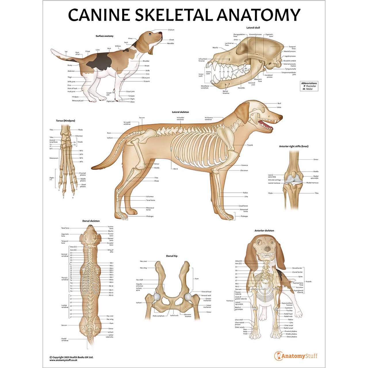
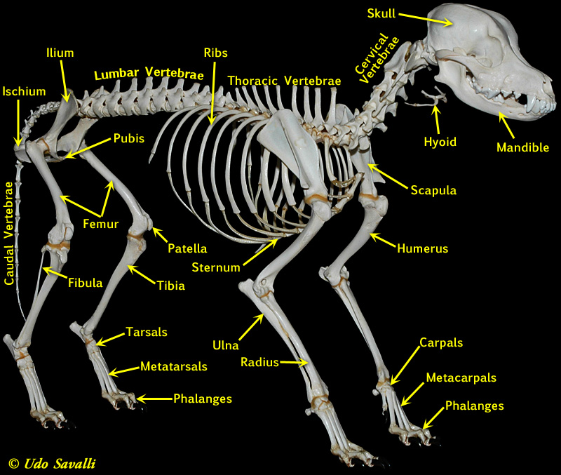
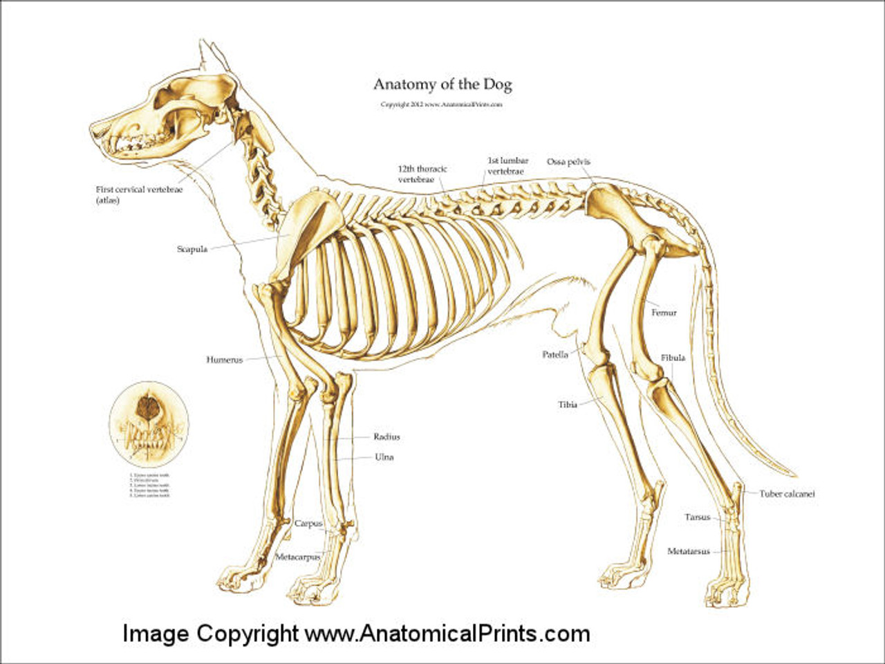
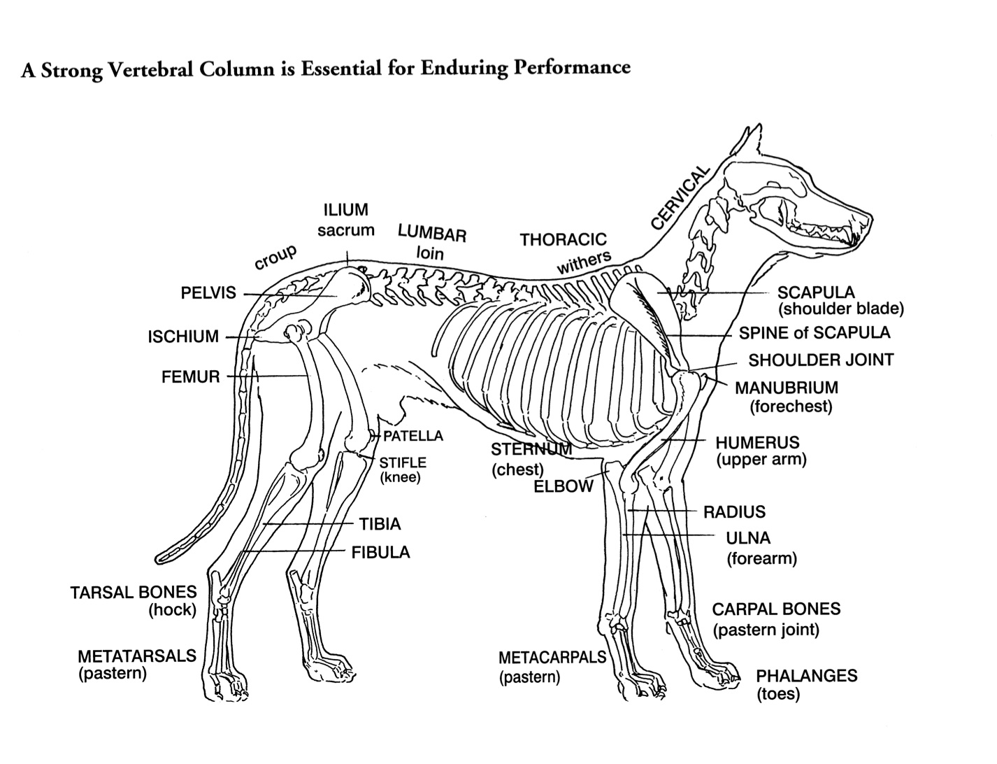
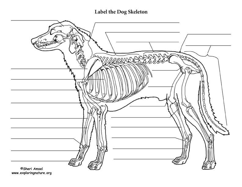
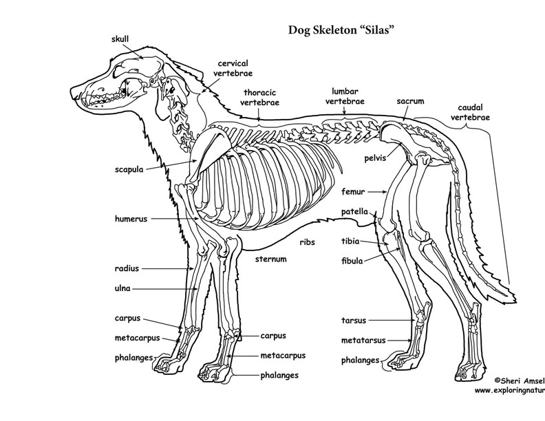
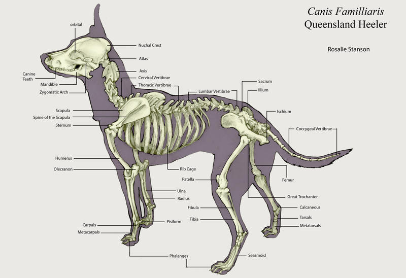
Dog skeleton. As with any vertebrate animal, the skeleton of a dog has the function of supporting the body for movement and protecting its internal organs. We can divide the canine skeleton into three main sections: Axial skeleton: skull, spine, ribs and sternum bones. Appendicular skeleton: bones of the extremities.
Canine Skeleton Poster Clinical Charts and Supplies
Speaking of skeletons, a dog has 320 bones in their body (depending on the length of their tail) and around 700 muscles. Muscles attach to bones via tendons. Depending on the breed of dog, they will have different types of muscle fibers. You've probably heard about slow and fast twitch muscle fibers before.
Helen King on Structure Evaluation Susan Garrett's Dog Training Blog
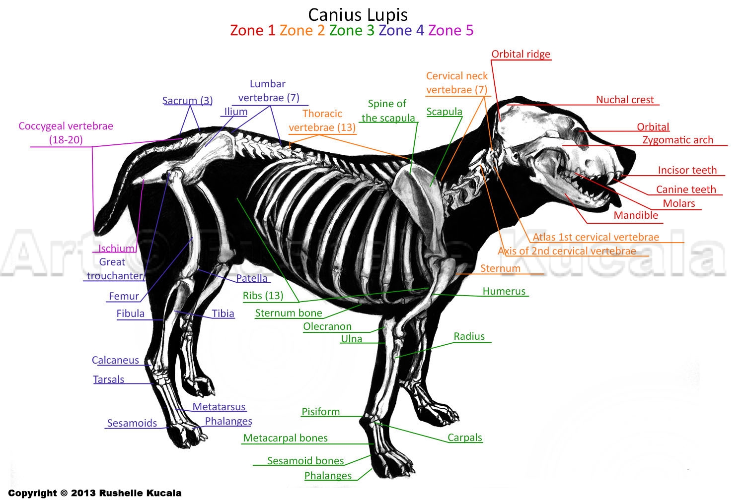
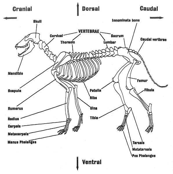
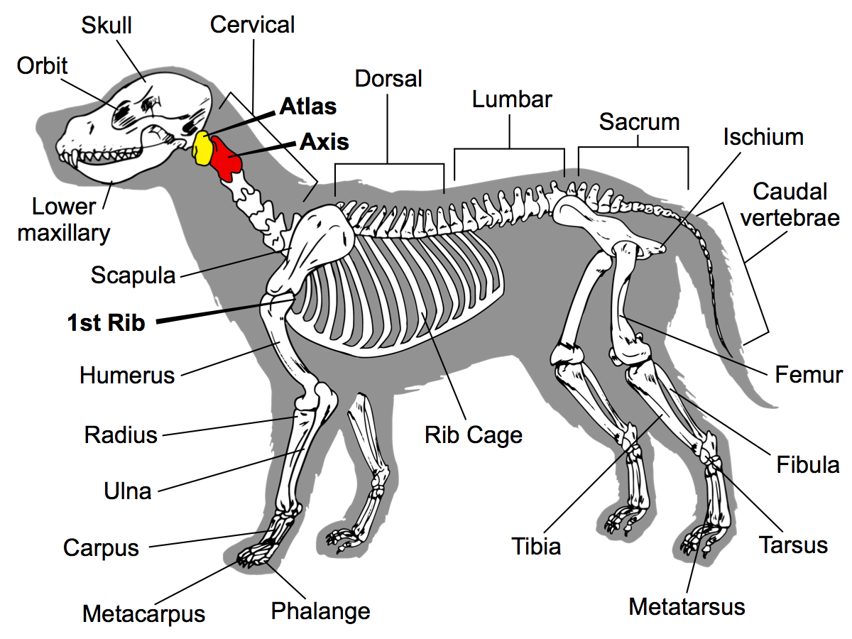
Here are presented scientific illustrations of the canine skeleton, with the main dog's bones and its structures displayed from different anatomical standard views (cranial, caudal, lateral, medial, dorsal, palmar..). Some of the different canine joints are labeled.
Dog Skeletal Anatomy
This module of vet-Anatomy presents an atlas of the anatomy of the head of the dog on a CT. Images are available in 3 different planes (transverse, sagittal and dorsal), with two kind of contrast (bone and soft tissues).
Dog Skeleton Anatomy by TheDragonofDoom on DeviantArt
25/04/2023 31/12/2021 by Sonnet Poddar The dog skeleton anatomy consists of bones, cartilages, and ligaments. You will find two different parts of the dog skeleton - axial and appendicular. Here, I will show you all the bones from the axial and appendicular skeleton with their special osteological features.
Types And Parts Of Bones Reading Ancient Animal Remains
Here, I will provide a dog skeleton labeled diagram and the different parts of a dog diagram. In the dog skeleton labeled diagram, I tried to show you all the bones from the body. This might help you understand the different regions of the body so quickly. I would like to show different external features of a dog again here in a labeled picture.
Home Study “Canine Musculoskeletal Unwinding” Watch Instantly Video
The forelimb skeleton consists of the thoracic or pectoral girdle and bones of the forelimb (see Figures 5-5 and 5-6). The size of forelimb bones varies a great deal, because of the greater variation in size for breeds of dogs. The forelimbs bear 60% of the dog's weight. The canine scapula is positioned close to the sagittal plane.
Dog Skeletal Anatomy
What are the main functions of the dog´s skeleton? The skeletal system provides stability and support to the muscles which in coordination with the muscular system, create movement. Another function is to protect the different parts of the body from possible blows or accidents.
Dog Bones labeled by Otvali on DeviantArt
Dog anatomy comprises the anatomical studies of the visible parts of the body of a domestic dog.Details of structures vary tremendously from breed to breed, more than in any other animal species, wild or domesticated, as dogs are highly variable in height and weight. The smallest known adult dog was a Yorkshire Terrier that stood only 6.3 cm (2.5 in) at the shoulder, 9.5 cm (3.7 in) in length.

Dog Skeleton Labeled Dog skeleton, Cat skeleton, Animal skeletons
Labeled atlas of anatomy: illustrations of the dog: Bones - Skeletal system Dog - Muscles Dog - Thorax/Abdomen/Pelvis Animal - Anatomy atlas: Cardiovascular system Veterinary anatomy - Animal: ANATOMICAL PARTS Abdomen Abdominal aorta Abdominal mammary gland Abdominal mammary region Accessory carpal bone Acromion Adductor muscle
